Melanoma Cells: 8K Scientifically Informed Illustration
Choose Your License
(Get better value with a credit pack)All sales are final. By purchasing, you agree to our Terms of Service.
This is an artistic interpretation for educational use. Please review our full Disclaimers before use.
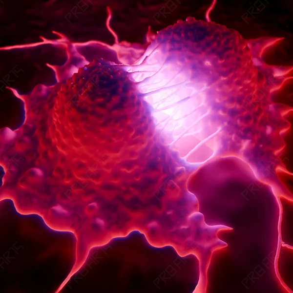
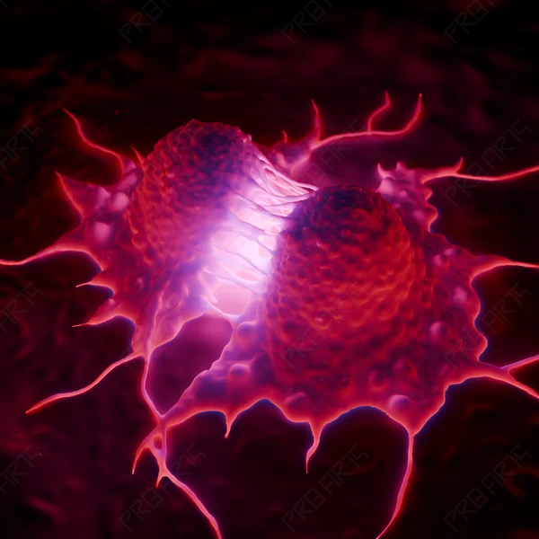
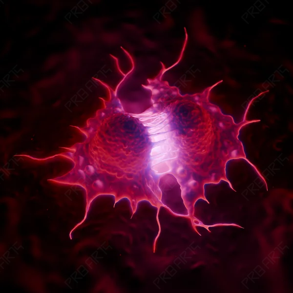
Experience the final, dramatic moment of cellular replication with this premium 8K visualization. This detailed conceptualization captures the precise instant of cytokinesis within a melanoma cell line, where the mother cell completes its transition into two distinct daughter cells. The striking contrast and cinematic lighting are designed to highlight the aggressive nature of malignant proliferation, making it a powerful visual for complex oncological narratives.
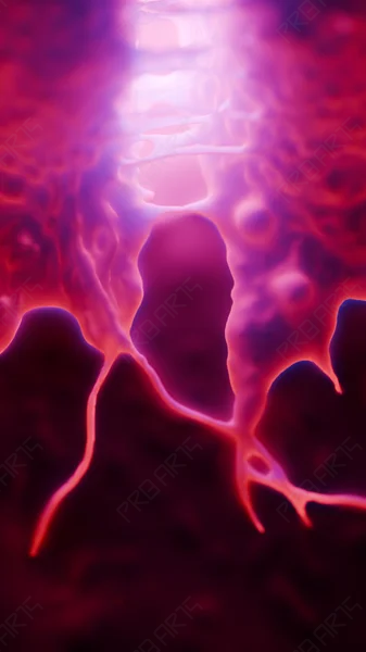
This asset bridges the gap between high-level science and cinematic art. Every filament and structural tension point is rendered with extreme clarity, providing an immersive look at the mechanics of cancer growth. It is an essential visual for professionals who require high-impact imagery that respects biological complexity without claiming clinical diagnostic accuracy.
🎥 Why This Asset Works:
-
8K Ultra-HD: Provides enough detail for large-scale medical exhibits and 8K broadcast displays.
-
Visual Tension: Captures the "final split," a specific stage often requested by educational publishers.
🎯 Perfect For:
-
Oncology researchers and pharmaceutical marketing teams.
-
Medical documentary filmmakers and high-end health journalists.
-
Educational institutions focusing on advanced cell biology and cancer pathology.
💡 Use Cases:
-
Keynote presentations for oncology conferences.
-
Hero imagery for pharmaceutical landing pages focusing on mitotic inhibitors.
-
High-definition educational video overlays and textbook covers.
🛠 Technical Specifications:
-
Resolution: 8K (8192 x 8192)
-
Format: High-Quality JPEG
-
Color Profile: sRGB / Adobe RGB optimized
🧬 Scientific Context: This illustration depicts the conclusion of the mitotic phase (M-phase) in a melanoma cell. It specifically visualizes the completion of the contractile ring and the physical separation of the cytoplasm, known as cytokinesis, resulting in two genetically identical malignant daughter cells.
- Asset ID
- 29
- Asset Type
- Illustration
- Dimensions
- 8192 x 8192 px